Plastic and Reconstructive Surgeon
Benign Moles
A naevus is a group of cells that contain melanin (brown) pigment. The pigment is usually found in a cell called a melanocyte. Depending on where the cells are located in the skin, the appearance of the naevus varies.
- A junctional naevus is smooth, flat and brown. They appear in childhood and occur on any site on the body. The melanocytes (pigment cells) are found at the junction of the epidermis and dermis.
- A compound naevus forms as the pigment cells descend into the dermis with increasing age. They occur in young adults. They are usually less than 5mm diameter, brown and slightly raised.
- An intradermal naevus is usually raised, pink or brown, with or without hair. The naevus cells are usually confined to the dermis. They usually occur in patients over 30 years of age, more commonly on the face.
- A Halo naevus is a brown mole surrounded by a "halo" of depigmented (white) skin. They frequently occur on the trunk of young adults. Over a few months the central pigmented naevus disappears leaving a pale area which may last for several years. The naevus resolves due to an intense immune reaction.
These naevi are benign and show no increased risk of malignant change to a melanoma over normal skin. Removal is indicated only if they change (size, shape, contour, texture), if symptoms develop (itch, pain, bleeding) or for cosmetic reasons.
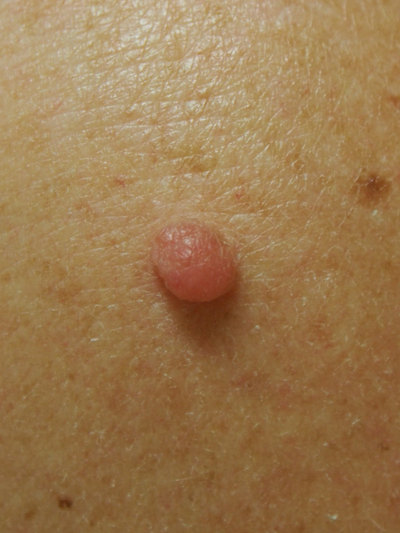
Intradermal Naevus
Dermatofibroma
These are firm, small (<1cm) pink to light brown nodules. They occur most commonly on the legs. They are lumps of scar tissue and may occur after an insect bite. Excision is only indicated if they are subject to repeated trauma (eg shaving), uncertain clinical diagnosis or for cosmesis.

Blue Naevi
Blue naevi are small well defined moles. The pigment cells lie deeply within the dermis giving them a blue-black colour.

Congenital Hairy Naevi
Congenitalhairy naevi are usually present at birth or soon after. They are flat brown moles in childhood but become raised, warty and hairy during teenage years. They vary in size from 1cm to the "Giant" variety which may cover large areas of the body. The giant variety has an increased risk of malignancy.

Dysplastic Naevi
Dysplastic naevi are usually larger than ordinary naevi, predominantly on the trunk and often show a mixture of tan, dark brown and pink areas. The surface texture may be pebbly and the border smudgy. The term “Dysplastic naevus syndrome” refers to the occurrence of multiple (>80) naevi on an individual. Melanoma risk is increased in these patients with the syndrome. The melanoma may arise in one of these abnormal naevi or in normal skin.

Seborrhoeic Keratoses (Warts)
Occur in adulthood and tend to be inherited. They start as a flat pink or brown patch and gradually become brown raised, greasy, warty growths. They increase in numbers with advancing age. They may grow up to several cm's across. Treatment is only indicated if there is any uncertainty in clinical diagnosis or for cosmesis.

Trichoepitheliom's
These are benign appendage tumours related to the hair glands in the skin. They usually occur after puberty and most commonly are found on the face. They are small pink nodules usually <3mm in size. Larger ones can be confused with skin cancers (Basal Cell Carcinoma) and may be excised to confirm diagnosis

Skin Tags
These are very common soft, skin coloured, round or pedunculated papilloma’s (polyp). They can get up to 1cm in size. They occur in middle to older age groups, and are more common in women or obese people. They mainly occur in the intertriginous areas (armpits, under the breasts, groin) and neck regions.
Vascular Lesions
- Strawberry Naevi (capillary haemangioma) is a soft, raised, bright-red to deep purple, vascular mark that develops at birth or soon afterwards and disappears spontaneously by the fifth year.
- Port-wine stains are irregularly shaped pink-purple flat marks in the skin that are present at birth but do not disappear with increasing age. Approximately 1% are inherited. The area of involvement tends to increase proportionally with the size of the child. In adulthood, they may become raised with nodular areas that may cause significant progressive cosmetic disfigurement. They can be treated with laser therapy.
- Venous lakes are dark blue to purple soft swellings occurring on the face, lips and ears in patients over 50 years of age. They result from a dilated (enlarged) vein. They blanch when pressure is applied to them (e.g. push on them with tip of pencil) and the blood is emptied out of them. They can be excised for cosmetic reasons.
Campbell de Morgan spots are common, often multiple, occurring in older individuals. They are formed by enlarged capillaries (small blood vessels) in the skin. They are named after an English surgeon, Campbell de Morgan (1811-76). The are flat to slightly raised cherry red - purple spots usually 2-3mm in size occurring principally on the trunk area but may occur anywhere on the skin. If removal is desired, treatment options include pulsed dye laser, electrocautery or excision.
Campbell De Morgan spots