Plastic and Reconstructive Surgeon
Plastic and Reconstructive Surgeon
CYSTS
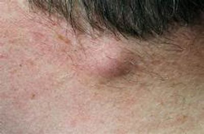
Sebaceous cysts arise from the glands in the skin. They are attached to the skin by the neck of the gland. This shows up on the skin surface as a small pinpoint, the punctum. Occasionally sebaceous material can be squeezed out of this hole. The bottom of the gland is expanded like a balloon, is lined by a thin layer of skin and contains sebaceous material which is white, greasy and smelly. They may grow up to 5cm in size. They can rupture or become infected (red, hot, swollen) like a boil.
Dermoid cysts These cysts are in the subcutaneous tissue. Congenital Dermoid Cysts present at or soon after birth and are often situated on the face around the eyes. They result from implantation of skin remnants along the lines of embryonic fusion of the segments of the face.
Inclusion Dermoid Cysts result from the traumatic implantation of a fragment of skin into the subcutaneous fat tissue. These cysts are lined by skin and filled with white sebaceous material.
Trichilemmal Cysts usually occur on the scalp in middle-aged women. They are often multiple and have a familial tendency. They arise in relation to hairs and are lined by a thick layer of skin and contain keratin (thick, cheesy material). They may grow up to 5cm in size.
Surgery
Most benign skin lesions can be excised under local anaesthetic in the consulting rooms unless they are very large, multiple or the patient cannot cope with a local anaesthetic e.g. children. The procedure takes 30-60 minutes. The only discomfort will be when the local anaesthetic is injected. This may sting for up to 1 minute. After this there's a 5-10 minute wait whilst the local anaesthetic numbs the region.
When excising a cyst a small ellipse of skin overlying the cyst including the neck of the blocked gland needs to be excised.
If your incision is on the face, the skin is usually stitched up with nylon sutures that will need to be removed 5 to 7 day's later. On other areas of the body dissolving sutures under the skin are usually used. The wound is covered when ever possible with a waterproof plastic patch so that you may shower. Strenuous exercise or work should be avoided for 7-14 days afterwards to minimize bleeding or splitting open the wound.
After one week you will be seen, the dressing will be changed and/or stitches removed. The lesion will have been sent for testing (histopathology) to check that it is benign (not malignant). You will be given the results at this appointment. The wound will usually be covered for another 2-7 days with a paper tape dressing.
Over the following months you need to massage the wound with a Silicone Scar gel or Bio-oil 1-2 times per day. For the first week massage gel / oil in very lightly to moisturize the wound. After that apply light pressure as you massage to help break up the scar tissue. We will usually arrange a time for you to return to the surgery after 4 to 6 weeks to check on how the scar is settling. At that time further scar management may be commenced such as silicone gel scar patches, ultrasound or a cortisone treatment to further improve your scar.
Possible Complications of Surgery
(1) Infection of the wound. Treatment with antibiotics may be necessary.
(2) Bleeding from the wound may require an early change of dressing, further treatment to stop the bleeding, or additional suturing.
(3) Haematoma or Seroma is a collection of blood or serum beneath the wound. It may require extra visits to drain the fluid or reopen the wound to allow removal of the blood and stop the bleeding.
(4) Wound breakdown; although the wound usually heals well in two weeks or so, complete healing may take weeks or months. Even after the stitches have been removed, strenuous activity or unexpected pulling may cause the wound to reopen. This can usually be avoided by ensuring that the area is not under physical stress for several weeks.
(5) Allergy to tapes; some patients develop an itchy rash from the dressing. This usually settles within a few hours to days of removing the dressing without permanent damage.
(6) Scarring depends on factors such as the size of the lesion, the rate of healing, your age and general health, and genetic make-up. Some people develop thick, raised, red and itchy hypertrophic scars. These are most common on the back, chest and shoulders. Over 1-2 years these scars improve, but they usually remain stretched or wide. Keloid scars are similar to hypertrophic scars but tend to grow beyond the edges of the original wound, producing a thick, raised lump bigger than the original wound. They may be painful or itchy. They occur more commonly in dark skinned races. They take many years to settle and often leave a poor scar. They can be treated with silicone gel patches, cortisone injections, and rarely may require re-operation with post operative radiotherapy.

LIPOMA
These are localised benign tumours composed of fat. They occur anywhere on the body, usually under the skin in the subcutaneous fatty tissue but may also involve deeper tissue including muscle. They slowly increase in size and may grow up to 30cm over many years. Familial Lipoma syndrome is an inherited (autosomal dominant) syndrome where in adulthood dozens of small non tender lipoma appear. Dercum’s disease usually occurs in middle aged women who develop multiple tender lipoma. Lipomas only need to be excised if there is any doubt regarding their diagnosis, if they are tender or if they are becoming a cosmetic problem.
Surgery
Most benign skin lesions can be excised under local anaesthetic in the consulting rooms unless they are very large (>3-4cm), multiple or the patient cannot cope with a local anaesthetic. If one of these, you would need to be admitted to hospital usually as a day case or one night stay. The procedure takes 30-60 minutes.
When under Local Anaesthetic the only discomfort will be when the local anaesthetic is injected. This may sting for up to 1 minute. After this there's a 5-10 minute wait whilst the local anaesthetic numbs the region. If done in hospital the procedure can be done under a general anaesthetic or local anaesthetic and sedation in some cases.
With a lipoma, no skin usually needs to be excised, with the incision being a little smaller than the width of the lipoma.
If your incision is on the face, the skin is usually stitched up with nylon sutures that will need to be removed 5 to 7 day's later. On other areas of the body dissolving sutures under the skin are usually used. The wound is covered when ever possible with a waterproof plastic patch so that you may shower. Strenuous exercise or work should be avoided for 7-14 days afterwards to minimize bleeding or splitting open the wound.
After one week you will be seen, the dressing will be changed and/or stitches removed. The lesion will have been sent for testing (histopathology) to check that it is benign (not a cancer). You will be given the results at this appointment. The wound will usually be covered for another 2-7 days with a paper tape dressing.
Over the following months you need to massage the wound with a scar gel such as Bio-oil or a silicone gel, 1-2 times per day. For the first week massage Bio-oil in very lightly to moisturize the wound. After that apply light pressure as you massage to help break up the scar tissue. We will usually arrange a time for you to return to the surgery after 4 to 6 weeks to check on how the scar is settling. At that time further scar management may be commenced such as silicone gel scar patches, ultrasound or a cortisone treatment to further improve your scar.
CHONDRODERMATITIS NODULARIS HELICIS
Chondrodermatitis Nodularis Helicis (CDNH) is a common, benign, painful condition usually affecting the helix (rim) or antihelix (outer rolled surface) of the ear. CDH more often affects middle-aged or elderly men, but about 30% of cases are seen in women.
The exact cause of CDH is unknown, although most authorities believe it is caused by prolonged and excessive pressure. Several anatomic features of the ear predispose persons to the development of this condition. The ear has relatively little subcutaneous tissue for insulation and padding, and only small blood vessels supply the skin and cartilage. Inflammation, swelling, and subsequent death of the skin or cartilage due to trauma, cold, sun damage, or pressure probably initiate the disease. In most cases, focal pressure on the stiff cartilage most likely produces damage to the cartilage and overlying skin. Sleeping on the affected side is usually an important etiologic factor.
CDH may occasionally be associated with autoimmune or connective-tissue disorders, including autoimmune thyroiditis, lupus erythematosus, dermatomyositis, and scleroderma. CDH occurs most commonly in fair-skinned individuals with severely sun-damaged skin; however, it can occur in persons of any races.
Presentation
The classic presentation of CDH is of a spontaneously appearing painful skin coloured nodule on the helix or antihelix surface of the ear. The nodule usually is less than a 1cm in diameter and has a slightly raised, rolled edge and central ulcer or crust. Onset may be precipitated by pressure, trauma, or cold. It is usually on the side the person sleeps on the majority of the time and is painfull when they do so.
It is important to have is reviewed by your doctor as sun spots and non melanoma skin cancers can appear very similar. They are usually more painfull than skin cancers. Biopsy is indicated if the diagnosis of CDH is in doubt.
Treatment
The primary goal should be to relieve or eliminate pressure at the site of the lesion. This is often difficult because of a preference or necessity to sleep on the side with the lesion. A pressure-relieving pad can be fashioned by cutting a hole from the center of a piece of foam, sleeping on a travel neck pillow with the ear in hole or by using a headband with a 1-2 cm thick piece of foam with a central hole (ear sized) cut from it to protect the ear. Alternatively, a special prefabricated pillow is available that helps relieve pressure on the ear..
Topical antibiotics may relieve pain caused by secondary infections. Topical and intralesional steroids also may be effective in relieving discomfort. If trauma, pressure necrosis, cold, or sun exposure is suspected as an exacerbating factor, then reduction of exposure is beneficial.
If specific efforts to relieve pressure are unsuccessful, surgical approaches almost always are needed.
Surgery
Various procedures have been used in the treatment of CDH. These procedures include wedge excision, curettage, electrocauterization, photodynamic therapy, carbon dioxide laser ablation, and excision of the involved skin and cartilage. In general, the recurrence rate is high unless the underlying focus of damaged cartilage is removed and the pressure relieved. Treatment involves removal of the damaged cartilage with a small area of overlying skin that is ulcerated. This is sent for pathology to confirm the diagnosis. The procedure is usually performed under local anesthesia in the rooms.
While surgical intervention is a mainstay of therapy, success is around 80-90%. On occasion multiple surgeries may be necessary as removal of underlying protuberant cartilage results in adjacent protuberances that can be site(s) of recurrence of CDH, owing to a change in pressure points.

This website uses cookies.
We use cookies to analyze website traffic and optimize your website experience. By accepting our use of cookies, your data will be aggregated with all other user data.